
Smile Design & Dental Photography: Minimal Gear, Maximum Predictability

Dental photography has evolved into an indispensable element of modern clinical practice, particularly in esthetic dentistry and smile design. Beyond documentation, clinical photography provides a universal language that connects the dentist, the patient, and the dental laboratory.
Evidence suggests that standardized pre-, intra-, and post-operative photographs improve diagnostic accuracy, increase patient acceptance of treatment plans, and facilitate precise communication with technicians. Yet, many clinicians hesitate to adopt routine photography due to the perceived complexity or cost of professional setups.
A Practical Protocol with Minimal Equipment
-
Camera: A DSLR with a macro lens is ideal, but even a high-end smartphone can suffice when paired with proper lighting.
-
Lighting: A simple ring flash or continuous LED light ensures consistent exposure and shadow control.
-
Essential Shots: At least eight standardized photographs—frontal, lateral, full smile, and occlusal views—form the baseline for analysis.
-
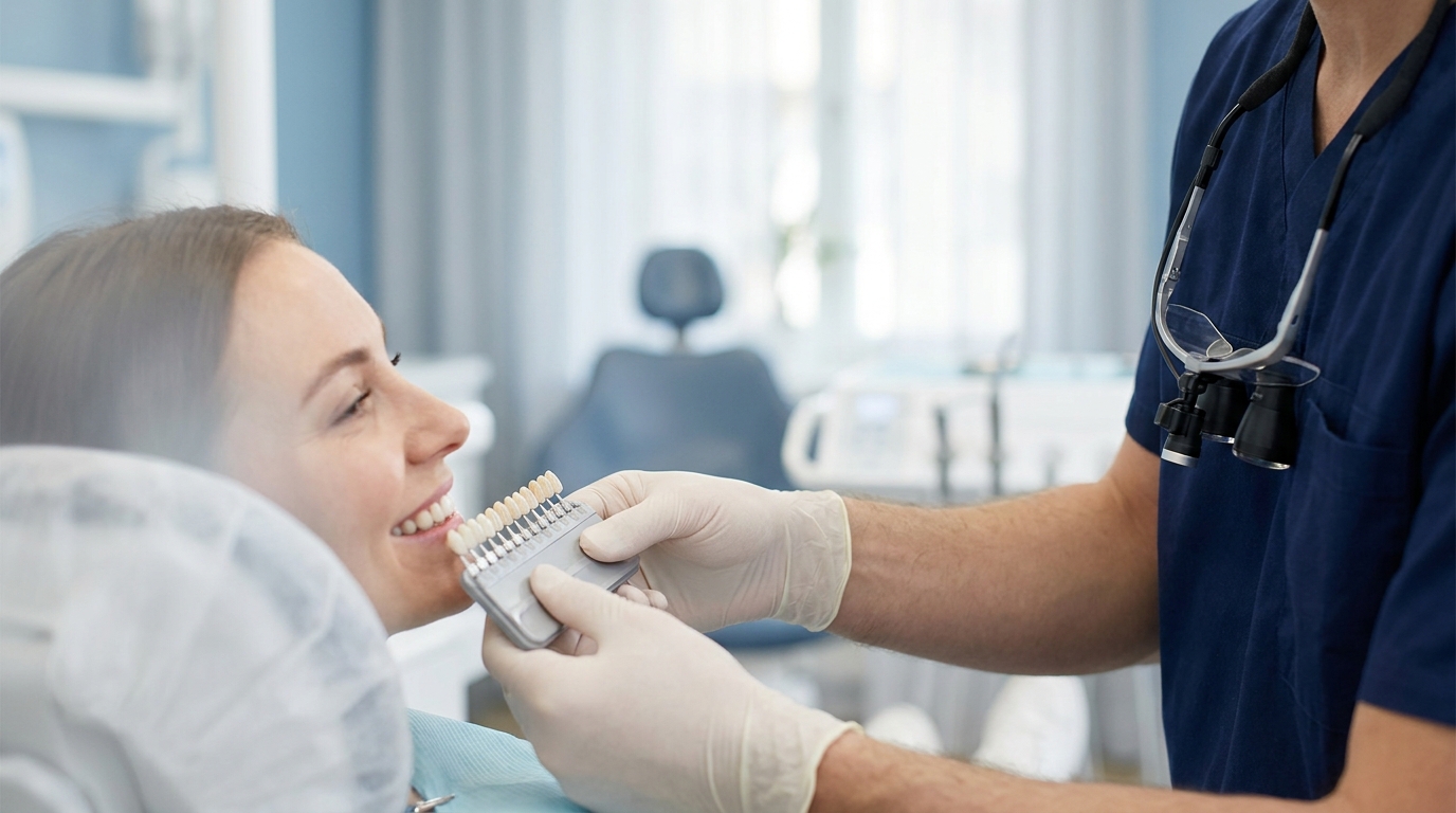
Color Calibration: A shade guide or gray card included in the frame helps achieve accurate color matching and communication with the lab.
-
Archiving: Organize photographs systematically (pre-/post-/intra-op), with clear file naming by patient and date for easy retrieval.
Advantages
-
Builds stronger trust with patients by visualizing expected outcomes.
-
Minimizes errors and miscommunication with the laboratory.
-
Enhances the educational and research value of each case.
Conclusion
Smile design does not require elaborate equipment to be predictable. By following a simple, standardized photographic protocol, clinicians can achieve high esthetic accuracy, improve workflow efficiency, and deliver consistent results that meet both functional and cosmetic expectations.